OS Trigonum Syndrome
Home / OS Trigonum Syndrome
OS Trigonum Syndrome
Get AppointmentWhat is OS trigonum syndrome (Posterior impingement)?
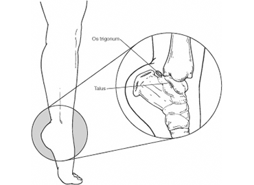
This condition is observed in athletes such as ballet dancers. The patient experiences pain at the posterolateral aspect of the ankle posterior to the peroneal tendon due to compression of the bone or soft tissue structures during activities that involve maximal ankle plantarflexion motion. It may also be seen in association with Flexor Hallucis Longus Tenosynovitis.
During the movement of plantar flexion, the foot and ankle are pointed maximally away from the body, the ankle is compressed posteriorly. This may result in tissue damage and pain if the compressive forces are too repetitive or forceful.
Posterior impingement often occurs due to inadequate rehabilitation following an acute ankle injury. In some cases, an individual may have an anatomical variant in their talus bone, known as an os trigonum, which is quite normal. However, such individuals have an increased likelihood of developing this condition, particularly in athletes.
Causes:
Pain might also occur if the flexor hallucis longus (FHL) tendon gets irritated. This can happen if the tendon doesn’t fit well because the tunnel is too tight or the tendon is too big, or if the tendon is inflamed and swollen (called tenosynovitis).
An ankle sprain may also cause a tear of the posterior ankle ligaments. The torn pieces can flip inside the joint. They can get pinched between the joint surfaces and cause pain. This problem is called posterior soft tissue impingement.
Posterior impingement syndrome can be triggered by an injury, such as an ankle sprain, or can occur chronically with activities or sports that frequently cause the downward pointing of the toes, such as ballet and gymnastics. The os trigonum or soft tissue can get squeezed between the ankle and heel bone.
Symptoms of OS trigonum syndrome
Individuals suffering from Posterior Ankle Impingement typically present with:
- Sharp pain at the back of the ankle joint during activities that require maximal plantar flexion (pointing).
- Ache at rest or following provocative activities.
Diagnosis
Posterior impingement syndrome can mimic other conditions such as an Achilles tendon injury, ankle sprain, or talus (ankle bone) fracture. Diagnosis begins with questions from your surgeon about the development of the symptoms and a thorough physical examination.
An X-Ray can be utilized when imaging posterior ankle impingement. The x-ray view of the ankle from the sideshows bone spurs. At times an x-ray taken at a slight angle (oblique radiograph) can be helpful in seeing anteromedial bone spurs.
MRI is a useful test for a couple of different reasons. First, it can be useful in being sure there is no other cause of foot or ankle pain present that can mimic posterior ankle impingement or additional symptoms.

Treatments
Relief of the symptoms is often achieved through treatments that can include a combination of the following:
- Rest. Limit the amount you are on the injured foot/ankle.
- Immobilization. A walking boot may be used to keep the foot and ankle from moving and allow the injury to heal.
- Ice. Apply an ice pack to the injured area, placing a thin towel between the ice and the skin.
- Compression. An elastic wrap may be recommended by your surgeon to control swelling.
- Elevation. The ankle should be raised slightly above the level of your heart to reduce swelling.
- Physical therapy. Your surgeon may start you on a rehabilitation program to help with the pain and to assist in function and strengthening.
- Medications. Nonsteroidal anti-inflammatory drugs (NSAIDs), may be recommended to reduce pain and inflammation. In some cases, prescription pain medications are needed to provide adequate relief.
- Injections. Sometimes cortisone and local anaesthetic are injected into the area to reduce the inflammation and pain.
Surgery Treatments
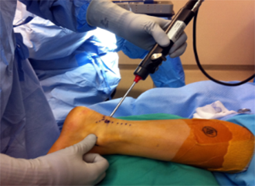
TArthroscopy is a surgical procedure in which the inside of a joint, such as the foot, is examined and treated using a camera and instrumentation that are inserted through small incisions typically one centimetre in length (called portals). Two small portals are typically used to perform the procedure.
Compared to traditional open incisions, the advantages of arthroscopy include:

- Earlier rehabilitation;
- Accelerated rehabilitation course;
- Less pain and blood loss;
- Lower rate of infection.
After surgery a splint or boot will be placed on the foot/ankle you had surgery on. You don’t put weight on the foot/ankle that was operated on until your surgeon tells you that you are allowed to. It usually takes eight to 12 weeks for athletes to return to play after posterior ankle arthroscopy and OS trigonum excision, but this time certainly can vary.
GET APPOINTMENT
Schedule Appointment : Your Path to Specialized Care
Get rid of your pain, stress, and enduring with our 24/7 dental services. It's a priority to relieve the pain in surgeon as much as possible. 90% of customers claim that they would come back & recommend us to others.
